What is Retinopathy of Prematurity?
Get Free Consultation
Chose Your Topic
What are the Symptoms of Retinopathy of Prematurity?
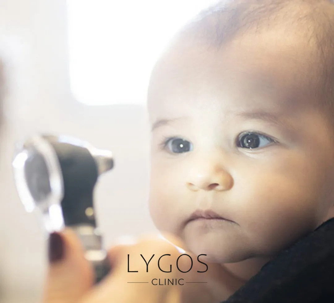
There are usually no obvious symptoms of retinopathy of prematurity that can be recognized by the patient or his/her relatives. This disease is monitored and diagnosed within the framework of special follow-up protocols determined for premature babies. ROP is detected by careful examination of the retinal layer behind the eye. Therefore, regular eye check-ups are vital for premature babies. Otherwise, the disease can progress in a way that is often not noticeable from the outside.

What are the Stages of Retinopathy of Prematurity?
ROP, Retinopathy of Prematurity, is classified into five different stages. These stages define the progression and severity of the disease. So, what are the stages of retinopathy of prematurity?
Stage I is characterized by mildly abnormal blood vessel growth. At this stage, the blood vessels are still developing normally, but this development is slightly impaired.
In stage II, the abnormal blood vessel growth is moderately increased. Most babies recover from these two stages without the need for treatment and regain normal vision. Sometimes the disease may go into remission on its own, but in other cases it may progress to Stage III.
In stage III, abnormal blood vessels begin to grow along the surface of the retina towards the center of the eye, not following normal lines of development. At this stage, some babies may recover without treatment, but if a certain level of what is known as “plus disease” develops, treatment may be needed. Plus disease is characterized by dilated and twisted blood vessels in the retina, a sign that the disease is getting worse. Intervening at this point increases the chances of preventing retinal detachment.
In stage IV, the scars caused by the bleeding cause the abnormal vessels to partially pull the retina away from the eye wall. This is the beginning of retinal detachment and can lead to vision loss.
Stage V is the final stage of the disease, in which the retina detaches completely and, if left untreated, the baby can develop severe visual impairment and even blindness.
While each stage determines the course of the disease and treatment requirements, early detection and appropriate intervention significantly improves babies' chances of preserving their eyesight.
How Does Retinopathy of Prematurity Occur?
When premature babies are born, the retina does not complete vascular development, which can lead to the formation of abnormal blood vessels in the non-vascular areas of the eye. This happens due to the effect of wrong signals (VEGF release).
These abnormal vessels can cause bleeding in the eye and detachment of the retina (retinal detachment), which can lead to severe vision loss. As birth weight and gestational age decrease, the risk of ROP (Retinopathy of Prematurity) increases. For example, while almost 80% of babies born at 25 weeks of gestation have ROP, this rate is around 10% in babies under 32 weeks.
This 10% group can usually recover spontaneously and do not require treatment. However, these babies should be checked regularly until vascular development is complete. ROP can occur not only in babies born under 1500 grams and before 32 weeks of gestation, but sometimes in babies born at later weeks (e.g. 36 weeks). If systemic risk factors are predominant in these babies, the neonatologist may still recommend ROP examination. However, all premature babies under 1500 grams and born before 32 weeks should be evaluated for ROP. These controls are of great importance for early diagnosis of possible complications and timely initiation of treatment.


How to Treat Retinopathy of Prematurity?
Early treatment is of great importance for babies diagnosed with Retinopathy of Prematurity (ROP). Various methods are used to prevent the progression of this disease. One of these treatment approaches is to deactivate the vascularized areas of the retina using laser beams.
This method prevents damage to healthy retinal tissue by areas that have not completed their development and preserves the baby’s existing vision. Another treatment option is the injection of drugs into the eye to remove the defective vessels. These injections stop the growth of the defective vessels and allow the retina’s vascular development to continue normally.
In this way, the intact retinal areas are preserved and the eye can continue to function. However, in some cases, more extensive surgical interventions may be needed due to a lack of early diagnosis or a very severe course of the disease. In such cases, surgical procedures are performed to minimize vision loss and improve the baby's quality of life. Although these procedures do not guarantee complete vision preservation, they are critical to maximize the vision of babies with ROP.

First Step
An eye examination is performed within 4-6 weeks after birth and the stage of ROP is determined.

Second Step
In the early stages, and especially in stage 3, laser treatment can be used to destroy abnormal blood vessels.

Step Three
It is an alternative to laser treatment. Cold is applied to stop the development of abnormal veins.

Step Four
In some cases, treatment with intraocular injections (e.g. anti-VEGF drugs) can be used.

Step Five
Babies' eyes should be monitored regularly after treatment.
Risk Factors for Retinopathy of Prematurity
Risk factors for retinopathy of prematurity play a critical role. These factors include low birth weight, presence of pre-eclampsia in the expectant mother, pulmonary hemorrhage in the infant, prolonged ventilation requirement and continuous positive pressure ventilation. Risk factors for retinopathy of prematurity may increase the progression and severity of ROP. Therefore, these infants should be monitored and treated more carefully. These findings call for early preventive measures and stricter follow-up of infants at risk.

Laser Eye Treatment in Premature Babies
In the treatment of ROP (Retinopathy of Prematurity), anti-VEGF injections may not be sufficient in some cases. In such cases, laser eye treatment can be used alone or in combination with injection therapy in premature babies. Which method will be used is determined by the evaluation of the condition of the baby’s eye.
Laser eye treatment in premature babies is usually performed under light sedation. In this procedure, the laser is applied to the non-vascularized areas of the retina to stop the function of the cells that cause the formation of false blood vessels in these areas. After the procedure, the baby’s condition is monitored at regular intervals. If the disease continues to progress, the laser treatment can be repeated or combined with anti-VEGF therapy.
However, if the disease progresses despite these treatment methods, surgical intervention may become inevitable. At this stage, surgery is used to preserve the baby's vision and prevent further damage. Early diagnosis and appropriate treatment are crucial to minimize vision loss in babies with ROP.
Eye Diseases in Premature Babies
Even after retinopathy of prematurity (ROP) has resolved, some people may still have eye problems. Premature babies may have more than one eye disease. These include lazy eye (amblyopia), refractive errors such as myopia and astigmatism, strabismus and glaucoma. In addition, tears may occur in the eye at an advanced age.
This can lead to vision loss. Therefore, children with ROP should closely monitor their eye health throughout their lives. In particular, regular eye examinations should be performed at least once a year and possible problems should be detected early. Treatment of eye diseases in premature babies is of critical importance. Early intervention can minimize the vision loss that these children may face in the following years.

Underdevelopment of Eye Vessels in Premature Babies
Get a quote for Retinopathy of Prematurity Treatment
Frequently Asked Questions About Retinopathy of Prematurity
BLOG

Is Breathing Through the Mouth Harmful?
Chose Your Topic Is Breathing Through the Mouth Harmful? Breathing is one of the most fundamental needs of life. However,

Does Rice Water Make Hair Grow? | Benefits of Rice Water
Chose Your Topic Does Rice Water Make Hair Grow? Natural methods in hair care have become quite popular in recent

Breast Lump | Types: Benign, Malign and Causes | LYGOS 2025
Breast Lump While cancer stands out as one of the most common health problems today, early diagnosis rates are also


